Improved Chronic Fatigue Symptoms after Removal of Mercury in Patient with Increased Mercury Concentration in Hair Toxic Mineral Assay: A Case
Article information
Abstract
Clinical manifestations of chronic exposure to organic mercury usually have a gradual onset. As the primary target is the nervous system, chronic mercury exposure can cause symptoms such as fatigue, weakness, headache, and poor recall and concentration. In severe cases chronic exposure leads to intellectual deterioration and neurologic abnormality. Recent outbreaks of bovine spongiform encephalopathy and pathogenic avian influenza have increased fish consumption in Korea. Methyl-mercury, a type of organic mercury, is present in higher than normal ranges in the general Korean population. When we examine a patient with chronic fatigue, we assess his/her methyl-mercury concentrations in the body if environmental exposure such as excessive fish consumption is suspected. In the current case, we learned the patient had consumed many slices of raw tuna and was initially diagnosed with chronic fatigue syndrome. Therefore, we suspected that he was exposured to methyl-mercury and that the mercury concentration in his hair would be below the poisoning level identified by World Health Organization but above the normal range according to hair toxic mineral assay. Our patient's toxic chronic fatigue symptoms improved after he was given mercury removal therapy, indicating that he was correctly diagnosed with chronic exposure to organic mercury.
INTRODUCTION
Fish contains essential ingredients and omega-3 fatty acid, and it is a nutritious food that is a part of a healthy daily diet. As a high-protein food, fish is especially recommended for young, pregnant, and old people. Some fish species, however, contain high concentrations of mercury that have accumulated through food chains.1) By consuming fish with concentrated mercury, people who are not exposed to mercury in their work environments can still experience chronic mercury exposure. Consuming significant amounts of fish can increase mercury concentrations in the blood, which offsets the nutritional gains from fish consumption.
Individual factors dictate mercury exposure symptoms differently.2) Chronic exposure to organic mercury mainly damages the nervous system, which causes fatigue, weakness, headaches, poor concentration, and emotional disturbance. Serious cases involve cognitive and sensory disorders, peripheral neuropathy, tremor, dysarthria, gait disturbance, visual disturbance, and auditory disorders.3) It has been reported that these symptoms can be alleviated by removing causative amalgams of mercury from patients with chronic mercury exposure who experience chronic fatigue, memory decay, and depression.4)
Generally, we confirm a diagnosis of idiopathic chronic fatigue when a patient passes the basic examination and chronic fatigue test; however, we continue to look for other plausible causes of symptoms as well. When the condition of a patient with fatigue does not improve with the usual treatment, we check the level of mercury in the patient's blood to determine if the level of mercury exposure is below the World Health Organization's specified criterion for confirming organic mercury intoxication but above normal levels. Here, we describe the case of a patient whose chronic fatigue improved after using medication to remove mercury from his body tissue.
CASE REPORT
A 47-year-old man who visited our department of family medicine, complained that he was experiencing chronic fatigue. His fatigue started about 1 year ago, and worsened with stress or after a day of work. His fatigue had previously improved after resting and sleeping but not for the last 6 months. He did not have a remarkable medical history, with the exception of treatment for reflux esophagitis. His dietary history included multivitamins and 4 weeks' consumption of red ginseng, 6 months ago. He did not smoke and consumed alcohol approximately twice a week. Additionally, he did not have any sleeping disorder of symptoms related to the digestive, nervous and circulatory systems, the results of a depression test were also normal, confirming that he was not suffering from any depressive disorder. Our patient's body temperature, heartbeat, and blood pressure were normal. No unusual signs were noted during simple neurologic testing or physical examination of the heart, lungs, abdomen, and muscle-bone system. There were no palpable lymph nodes in his neck, axilla, and perineum. The results of a peripheral blood test conducted to diagnose chronic fatigue were also normal as follows: hemoglobin, 15.6 mm Hg; platelets, 267/el; white blood cells, 7,390/bl; and erythrocyte sedimentation rate, 3 mm/h. In serum chemistry testing, the following levels were noted: aspartate aminotransferase, 34 IU/L; alanine aminotransferase, 35 IU/L; total protein/albumin, 7.5/4.8 g/dL; globulin, 2.6 g/dL; alkaline phosphatase, 182 IU/L; Ca/P, 9.9/3.9 mg/dL; fasting plasma glucose, 71 mg/dL; blood urea nitrogen, 11.8 mg/dL; creatinine, 0.65 mg/dL; sodium, 150 mEq/L; potassium, 4.1 mEq/L; and thyroid-stimulating hormone, 3.6 id. The routine urine analysis results were also normal. Antinuclear antibody and acquired immune deficiency syndrome test results were negative, and no specific signs were observed on abdomen ultrasonography and colonoscopy. However, we observed chronic gastritis and reflux esophagitis during gastroscopy. We recommended aerobic exercise, provided supportive psychotherapy, and prescribed medication for reflux esophagitis. We also asked the patient to submit a dietary diary and visit us again after 1 month. One month later, the patient said he had experienced no improvement in his fatigue and that he was extremely fatigued after these workouts as well. His dietary diary revealed that he ate slices of raw tuna more than twice per week. We eventually learned that raw tuna was his favorite food and that he has been consuming it for the past 5 years, partly because of his occupation. Suspecting methyl-mercury exposure, we conducted a blood test. We also conducted the hair toxic mineral assay (HTMA), after confirming that he had not permed, dyed, or coated his hair or used any functional shampoos. Mercury levels from blood and urine tests were within normal limits. We subsequently administered the patient a dietary medication designed to remove heavy metals. Each capsule contains zinc oxide, magnesium oxide, calcium, and L-cysteine.
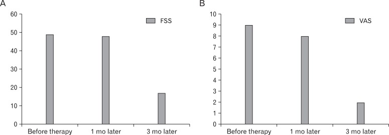
After administering a dietary medication designed to remove heavy metals for 3 months, we found that the mercury concentration in our patient had decreased to 1.972. Using the fatigue severity scale (FSS) and visual analogue scale (VAS) (Figure 1), we measured the patient's degree of fatigue before and after proem intake for 3 consecutive months.5) His FSS score was 49, and his VAS was 9 before Proem intake. One month after Proem administration, his FSS score was 48 and VAS was 8. After 3 months of intake, his FSS score reduced to 17 and his VAS reduced to 2 (Figure 2).
DISCUSSION
In the 1994 Centers for Disease Control and Prevention definition of chronic fatigue syndrome, the agency stated that although many diseases need to be ruled out when attempting to diagnose chronic fatigue, chronic fatigue syndrome should be diagnosed if a detailed clinical assessment shows that symptoms conform to a chronic fatigue definition even if chronic fatigue syndrome criterion are not met.6) According to reports, the causes of chronic fatigue can be identified in two-thirds of patients with chronic fatigue; 46% of these patients were diagnosed with characteristic organic diseases related to fatigue.7)
Fish consumption continues to increase in Korea as people avoid consuming meat due to the mad cow disease crisis and avian influenza; people assume that fish is safer than other meats. This increased consumption implies that the dangers of methyl-mercury accumulation and toxicity may be increasing. Therefore, as suggested in the educational reports on the diagnosis and treatment of chronic fatigue, any possibility of exposure to toxic materials must be explored.8)
Mercury can be categorized as elemental, inorganic, and organic.9) Unlike elemental and inorganic mercury, which mainly are absorbed through the lungs and skin, 95% of organic mercury is quickly absorbed by the digestive organs. Phenyl mercury, one type of organic mercury, is usually contained in spermicides and fungicides, but these compounds are no longer used. Ethyl-mercury, another type of organic mercury, is found in thimerosal, a vaccine preservative, but the extent to which thimerosal can harm humans remains controversial. Methyl-mercury, the most common organic mercury, is accumulated through the food chains of the ecosystem. It is found in fish, crustaceans, and marine mammals and is more highly concentrated in carnivorous fish with longer life spans. Consumption of seafood such as fish is the primary source of exposure to organic mercury.10-13)
Absorbed methyl-mercury is conjugated with cysteine, which is an amino acid that is abundant in inner-body proteins. The methyl mercury-cysteine conjugation penetrates the inside of a cell through the amino-acid transporter and is accumulated there. It also easily travels through the blood-brain barrier. Consequently, the conjugation is oxygenated and accumulated, which is toxic to humans and is called the chronic exposure of methyl-mercury.14-16) Symptoms resulting from chronic exposure to methyl-mercury gradually develop and may be observed after a long period of time. The main damage occurs in the nervous system and symptoms include fatigue, decay, headache, poor concentration, and mental disorders.17)
Among mercury-exposure diagnostic methods, blood tests are useful to detect acute exposure; urine tests, to detect chronic exposure to elemental or inorganic mercury; and HTMA, to detect chronic exposure to organic mercury (methyl-mercury).3)
The first priority when treating mercury exposure is to block source of exposure to organic mercury. In the case of acute exposure, a chelating agent can be directly injected into the blood. However, in cases of chronic exposure, because mercury has infiltrated the organs rather than the blood, it is helpful to promote a synthetic reaction within the organs which can chelate the heavy metal. Metallothionein is one such material that and removes heavy metals, and its synthesis is promoted by zinc.18) In our patient, we administered specific heavy metal removal medication containing zinc and cysteine to chelate and remove the mercury.
A research series reported a positive connection between the concentration level of methyl-mercury in the hair and frequency of fish consumption.19,20) However, whether a low level of methyl-mercury exposure from fish consumption is toxic to adults remains controversial.21) In a study of pregnant women and fetuses with low levels of mercury exposure in the blood, methyl-mercury was found to infiltrate the placenta and blood-brain barrier and caused developmental disorders in fetal brains and nerves, but the exposure presented almost no effect in the pregnant women.22,23)
Early symptoms of nerve intoxication from organic mercury exposure manifest at the level of 200 µg/L in a blood test, and 50 µg/g in a hair assay.24-26) In natives residing near the Amazon River, malfunction of the nervous system was observed at levels lower than intoxication from organic mercury exposure.27,28) In a study that considers the effect of low-level methyl mercury exposure on adult nervous-mental functions, the average mercury concentration of the subjects was 4.2 ± 2.4 µg/g. Depending upon the dose, mercury exposure caused distinctive changes in fine motor skill, dexterity, and concentration. Specific differences were found in fine motor speed (3.6 µg/g), digit symbol (3.8 µg/g), total logical memory (3.7 µg/g), backward digit span (3.6 µg/g), easy learning (3.7 µg/g), and the logical memory first-story test (3.7 µg/g).29) In 2008, the World Health Organization provided a criterion to measure the dangers of methyl-mercury by fish consumption; the criterion was that mercury concentration exceeding 2 µg/g in the hair can be dangerous.30) Likewise, many studies have been conducted to determine the accumulation of methyl-mercury in amounts lower than 50 µg/g.
It cannot be concluded that all chronic low-level organic mercury exposure (mercury concentration levels between 2 and 50 µg/g in the hair) will cause fatigue. Because it is difficult to identify factors that cause chronic fatigue other than low-level exposure to organic mercury via fish consumption, and because there is no response to conventional treatments for chronic fatigue, we believe that low-level exposure to organic mercury can cause chronic fatigue. It is difficult to conclude a causal relationship from our study as we have only described a single patient here. However, as our treatment was effective, it is clear that chronic idiopathic fatigue symptoms can improve after removing mercury from the body.
ACKNOWLEDGMENTS
This work was supported by research grant of the Wonkwang University in 2009.
Notes
No potential conflict of interest relevant to this article was reported.

