Association between Body Fat and Bone Mineral Density in Normal-Weight Middle-Aged Koreans
Article information
Abstract
Background
Osteoporosis and osteopenia are characterized by reduced bone mineral density (BMD) and increased fracture risk. Although the risk of fractures is higher in underweight people than in overweight people, the accumulation of body fat (especially abdominal fat) can increase the risk of bone loss. This study aimed to evaluate the association between body fat percentage and BMD in normal-weight middle-aged Koreans.
Methods
This study included 1,992 adults (mean age, 48.7 years; 52.9% women). BMD and body fat were measured using dual-energy X-ray absorptiometry. Multiple linear regression analyses and analysis of covariance were used to assess the association between BMD and body fat. Body fat percentage was grouped by cut-off values. The cut-off values were 20.6% and 25.7% for men with a body mass index of 18.5–22.9 kg/m2, while the cut-off values were 33.4% and 36% for women.
Results
Body fat percentage tended to be negatively associated with BMD. Increased body fat percentage was associated with reduced BMD in normal-weight middle-aged adults. The effects of body fat percentage on BMD in normal-weight individuals were more pronounced in men than in women.
Conclusion
There was a negative correlation between BMD and body fat percentage in middle-aged Korean men and women with normal body weight. This association was stronger in men than in women.
INTRODUCTION
Osteoporosis and osteopenia are characterized by reduced bone mineral density (BMD) and increased fracture risk. Bone strength is determined by bone mass and quality, and reduced bone strength increases the risk of fracture. Bone mass is mainly determined by BMD, which is usually measured to diagnose osteoporosis.
Several studies have shown that being underweight is a risk factor for fractures. However, previous studies have also shown that the mechanical loading of body weight onto the bone stimulates bone formation, resulting in increased BMD [1,2]. While some studies have simply identified weight as a variable, it is necessary to consider body fat and lean body mass separately in order to determine the relationship between body weight and BMD. One study has shown that fat accumulation, especially abdominal fat, acts as a risk factor for bone loss [3]. In a study of the relationship between body fat percentage and BMD, it was reported that a higher body fat percentage, regardless of body weight, was associated with an increased risk of osteoporosis and fracture [4]. One study also reported that high lean body mass and low fat mass have a protective effect on bone health in Korean men [5].
Although many studies have examined body fat percentage and BMD, their conclusions have been controversial, and few studies have focused on body fat percentage independently from other variables. In normal-weight individuals, it is possible to focus on the relationship between body fat percentage and BMD by reducing the influence of various other factors related to the presence of excess fat that affect BMD, such as hormones, cytokines, and mechanical loading. Accordingly, the current study used data collected by the Korea National Health and Nutrition Examination Survey (KNHANES) from 2009–2011, which suggested cut-off values for the increased risk of cardiovascular disease in order to distinguish normal-weight obese participants from normal-weight participants with a 18.5 kg/m2≤ body mass index (BMI) <23.0 kg/m2. The present study grouped participants based on these values6) and aimed to determine the relationship between body fat percentage and BMD in normal-weight individuals.
METHODS
1. Sample Population
This study used raw data from the 4th (2008, 2009) and 5th years (2010, 2011) of the KNHANES published by the Korea Centers for Disease Control and Prevention of the Ministry of Health and Welfare [7]. The National Health and Nutrition Survey is conducted every year to identify the health and nutritional status of the general population and is currently being released every 3 years. The 7th (2016–2018) survey is currently in progress.
Among the 37,753 participants surveyed, 12,740 were men and women aged 40 to <65 years; among these, 4,284 (1,612 men and 2,672 women) had a 18.5 kg/m2≤ BMI <23.0 kg/m2 (the normal-weight group). People with rheumatoid arthritis, renal failure, diabetes, thyroid disease, hepatitis, liver cirrhosis, and cancer were excluded. Furthermore, people who received osteoporosis treatment, pulmonary tuberculosis treatment, oral contraceptives, and female hormone therapy were excluded, and a total of 1,992 people (938 men and 1,054 women) were included in the analysis. Only those who underwent both BMD and body composition tests were included. The KNHANES was approved by the institutional review board of the Centers for Disease Control and Prevention(2007-02CON-04-P, 2008-04EXP-01-C, 2009-01CON-03-2C, 2010-02CON-21-C, 2011-02CON-06-C) from 2007 to 2011.
2. Body Measurement
Anthropometric measurements of the entire body were performed by trained investigators. Height and weight were measured to the nearest 0.1 kg and 0.1 cm, respectively, with the participant barefoot. BMI was calculated by dividing body weight (kg) by the square of height (m2). Waist circumference was measured to an accuracy of 0.1 cm while keeping the circumference of the middle portion of the body (between the bottom of the last frame and the top of the iliac crest) in an upright state.
Dual-energy X-ray absorptiometry was used to assess BMD and body composition. The BMD of the femur, femur neck, lumbar spine, and whole body were used, and body fat percentage was calculated by dividing the total fat mass by the total body weight and multiplying by 100.
3. Lifestyle
People were asked about their smoking, drinking, and physical activity habits through questionnaires. Smoking status was classified either as non-smoking, past smoking, or current smoking. The non-smoking category included people with no smoking experience or a history of smoking <100 cigarettes; past smokers include those who smoked >100 cigarettes in the past but no longer smoked; current smokers included those who currently smoked >100 cigarettes per year.
Drinking status was also classified into non-drinkers, mild to moderate drinkers, and heavy drinkers. Non-drinkers were those who had no experience of drinking alcohol, mild to moderate drinkers were those who drank <30 g/d (men) or <20 g/d (women), and heavy drinkers were those who drank >30 g/d (men) or >20 g/d (women).
Participants were classified into 3 groups according to their degree of physical activity, including inactive (0–599 MET-min/week), mild (600–2,999 MET-min/week), and intense (≥3,000 MET-min/week).
4. Statistical Analysis
All results were expressed as means±standard error and analyzed using IBM SPSS ver. 22.0 (IBM Corp., Armonk, NY, USA). Since the sample included in the KNHANES was obtained using the composite sample design method, a composite sample analysis was conducted. Missing values were treated as valid to exclude the result that the standard error of the estimate was biased.
We used the cut-off values for normal-weight obesity (18.5 kg/m2≤ BMI <23.0 kg/m2) published in a previous study of adults aged ≥20 years. The study population was divided into three groups based on the body fat percentage cut-off values (20.6% and 25.7% for men and 33.4% and 36.0% for women) [6].
Analysis of variance and the chi-square test were used to compare the general characteristics of enrollees. Multivariate linear regression analysis and analysis of covariance (ANCOVA) were performed to examine the association between total hip, femoral neck, lumbar spine, and total BMD in the body fat percentage groups and gender groups. The data were corrected for age, BMI, smoking, drinking, and physical activity, and the presence of menopause was taken into consideration in women.
RESULTS
1. General Characteristics
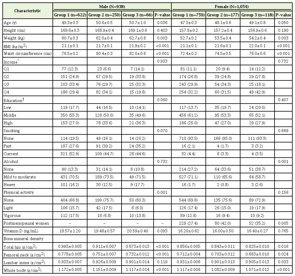
Table 1 shows the general characteristics of the study population and the differences between groups and men and women. Among the men, 622 participants (66.3%) were in group 1 (normal, total body fat <20.6%), 250 participants (26.7%) were in group 2 (overweight, 20.6%≤ total body fat <25.7%), and 66 participants (7.0%) were in group 3 (obesity, total body fat ≥25.7%). Among the women, 759 (72.0%) were in group 1 (normal, total body fat <33.4%), 177 (16.8%) were in group 2 (overweight, 33.4%≤ total body fat <36.0%), and 118 (11.2%) were in group 3 (obesity, total body fat ≥36.0%).
There were no statistically significant differences in educational level, income, smoking, and alcohol intake between men and women. There was a significant difference between groups in terms of physical activity and age in men, and no differences between men and women. There were statistically significant differences in body weight, BMI, and waist circumference between the groups, although there was no statistically significant difference in height between the groups in both men and women. In the case of women, there was a statistically significant difference in the percentage of postmenopausal women between the groups. Total hip, femoral neck, lumbar spine, and total BMD showed a tendency to decrease as the total body fat percentage increased, and there was a statistically significant difference between the groups except in the lumbar spine in men.
2. The Relationship between Bone Mineral Density and Body Fat Percentage
Table 2 shows the estimated mean values of BMD according to body fat percentage obtained through ANCOVA. Model 1 was adjusted for age only, while model 2 was added to model 1 to adjust for BMI. In addition to model 2, model 3 was adjusted for smoking, drinking, and physical activity. In women, menopause was taken into consideration.

Analysis of variance for the bone mineral density of the total hip, femoral neck, lumbar spine, and whole body
Estimated mean values for both men and women showed a tendency to decrease from group 1 to group 3 except in the lumbar spine. Female lumbar spine BMD decreased from group 1 to group 2 but increased in group 3. In the male lumbar spine, female femur neck, and female lumbar spine, the trend was not statistically significant in model 1. However, after additional adjustment, the results showed that the total hip, femoral neck, lumbar spine, and whole body BMD decreased significantly with increasing body fat.
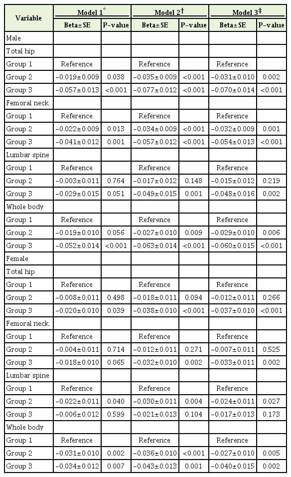
Table 3 shows the effect of BMD according to body fat percentage via multivariate linear regression analysis. Groups 2 and 3 were compared and analyzed with respect to group 1. First, the total hip and femur neck BMD of men from group 1 versus group 2 and group 2 versus group 3 showed statistically significant results in all models. Total body BMD showed statistically significant results except in group 1 versus group 2 in model 1. For lumbar spine BMD, even after gradual correction, the group 1 versus group 2 comparison showed no statistically significance, while the group 1 versus group 3 comparison showed a statistically significant difference.

Multiple linear regression analyses for the bone mineral density of the total hip, femoral neck, lumbar spine, and whole body
In women, the difference in the BMD of the entire femur and femoral neck between group 1 and group 2 was statistically non-significant after calibration, but the same differences between group 1 and group 3 were statistically significant.
DISCUSSION
The factors affecting BMD vary widely, and their interactions determine its level. It is well known that lifestyle and eating habits are important and that aging itself also reduces bone mass. Several studies have shown that obesity has a negative effect on BMD, and the mechanism for this is multifactorial. Some studies have shown that the body experiences increases in inflammatory cytokine levels during obesity and that these changes are related to bone loss [8]. Studies in Korea have also shown that visceral adiposity is inversely related to BMD [9] and that visceral adipose tissue has a deleterious effect on the lumbar micro-architecture in postmenopausal women. Subcutaneous adipose tissue has beneficial effects on BMD [10]. However, few studies have investigated the adipokines secreted from these fat tissues.
The results of this study are not significantly different from the results of previous studies in which body fat percentage was found to be negatively correlated with BMD [4,11]. Interestingly, lean body mass is actually positively correlated with BMD [5]. In the present study, we used the cut-off values presented in a previous study to categorize individuals and explore the relationship between body fat percentage and BMD. Therefore, unlike previous studies, the influence of other factors that may affect BMD was minimized, and we contend that the negative effect of body fat percentage on BMD is due to fat tissue itself.
Overall, the decrease in BMD with increasing body fat percentage was less significant in women than in men. In the lumbar spine, the effect of body fat percentage reduction was less significant than that in the whole body, total hip, and femoral neck. In the lumbar spine (especially in women), BMD increased as a result of increased body fat percentage. Previous studies have reported that the volumetric BMD of the lumbar spine is related to fat mass, not lean body mass [12], and one study reported a positive association between trunk fat and spinal BMD in postmenopausal women [13]. However, the underlying mechanism of this relationship is unclear, and further research is warranted.
This study is based on the large scale data of the KNHANES, which allowed us to include a large number of participants. In addition, the association of BMD with increased body fat percentage was confirmed in the normal-weight group (18.5 kg/m2≤ BMI <23.0 kg/m2). This study is the first to demonstrate the concept of normal-weight obesity by comparing and analyzing BMD according to subgroups of obesity-related risk cardiovascular risk factors.
The first limitation of this study is its cross-sectional design, which made it difficult to confirm a causal relationship between body fat percentage and BMD, although an association was detected. Therefore, it is not clear whether increased body fat percentage directly leads to a decrease in BMD, and further prospective studies are needed. Second, the relationship between body fat and BMD was analyzed according to body fat percentage only, and the relationships between lean mass, abdominal obesity, or visceral obesity and BMD were not investigated. However, considering that the difference in height between the groups was not statistically significant and the waist circumference increased significantly from group 1 to group 3, the increase in body fat percentage in the normal-weight group was mainly due to an increase in abdominal fat. Third, the presence of menopause was corrected for. However, since the exact timing of menopause was not investigated, we were unable to evaluate BMD according to menopause. Finally, we did not investigate correlations between BMD and calcium intake, serum vitamin D, and serum estrogen levels.
In conclusion, there is a negative correlation between BMD and body fat percentage in middle-aged Korean men and women with normal body weight. This association was stronger in men than in women.
Notes
No potential conflict of interest relevant to this article was reported.

