Usefulness of Pep-Test for Laryngo-Pharyngeal Reflux: A Pilot Study in Primary Care
Article information
Abstract
Background
Gastroesophageal reflux disease is a digestive disorder characterized by nausea, regurgitation, and heartburn. Gastroesophageal reflux is the primary cause of laryngeal symptoms, especially chronic posterior laryngitis. The best diagnostic test for this disease is esophageal impedance-pH monitoring; however, it is poorly employed owing to its high cost and invasiveness. Salivary pepsin measured using a lateral flow device (Pep-test) has been suggested as an indirect marker of laryngopharyngeal reflux (LPR). The present study tested the reliability of Pep-test in diagnosing LPR in uninvestigated primary care attenders presenting with chronic laryngeal symptoms, and evaluated the raw pepsin concentration in patients with LPR.
Methods
A multicenter, non-interventional pilot study was conducted on 86 suspected patients with LPR and 59 asymptomatic subjects as controls in three Italian primary care settings. A reflux symptom index questionnaire was used to differentiate patients with LPR (score >13) from controls (score <5). Two saliva samples were collected, and comparisons between the groups were performed using two-sided statistical tests, according to variable distributions.
Results
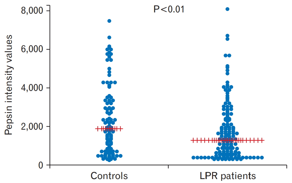
There was no statistical difference in the salivary pepsin positivity between LPR patients and controls, whereas the pepsin intensity value was higher in controls than in LPR patients.
Conclusion
A high prevalence of pepsin positivity was observed in asymptomatic controls. Pepsin measurement should not be considered as a diagnostic test for LPR in primary care patients.
INTRODUCTION
Laryngopharyngeal reflux (LPR) refers to the backflow of gastric contents into the larynx, pharynx, trachea, and bronchus, resulting in several upper airway inflammatory disorders. LPR is associated with several clinical manifestations, such as laryngitis, obstructive sleep apnea, posterior glottis edema and erythema, laryngospasm, subglottic stenosis, and even otitis media [1,2]. Patients with LPR generally show no specific symptoms, making the diagnosis difficult [3].
Gastroesophageal reflux disease (GERD) is a gastric disorder and probably one of the main causes of laryngeal symptoms, especially in patients with chronic posterior laryngitis. GERD may contribute to extraesophageal syndromes via both direct (aspiration of liquid and aerosol) and indirect (vagal mediated) mechanisms (sensory neuropathic cough) [4].
Data on the prevalence of LPR have remained controversial, with certain studies reporting it to range from 18% to 80% [1,5], whereas others reporting it as low as 10% [6]. According to a study, LPR is common in 26% of patients presenting to their general practitioners (GPs) with suspected symptom profiles [7]. However, establishing an association between GERD and symptoms of laryngeal irritation is challenging. The currently used diagnostic techniques for LPR, which rely on procedures having normative standards established for the diagnosis of typical GERD (i.e., heartburn and/or regurgitation), are not appropriate for the detection of LPR.
According to the current guidelines on GERD management, all patients presenting otolaryngological and respiratory disorders should be carefully evaluated for non-GERD causes [8]. A proton pump inhibitor (PPI) test is recommended to treat extraesophageal symptoms, particularly in patients who also have typical GERD manifestations. Although a recent meta-analysis showed that a high-dose of PPIs was no longer effective in improving LPR symptoms, high dosage and long duration of PPI therapy are required for the diagnosis of atypical GERD presentation [9]. Reflux monitoring should be considered before starting PPI therapy in patients with extraesophageal manifestations, who do not have typical GERD symptoms or do not respond to PPIs. However, it is not an ideal diagnostic tool because of low sensitivity (50%–80%), low patient tolerance, major diet influence, high cost, and limited availability [10,11].
In clinical practice, primary care patients with chronic laryngeal symptoms are often referred to otolaryngologists, who frequently establish a diagnosis of posterior laryngitis by laryngoscopy and prescribe PPIs and an upper digestive endoscopy to confirm the diagnosis. However, this is a non-rationale, non-evidence-based, expensive, and ineffective approach. Therefore, a less expensive and easier to perform surrogate marker of extraesophageal reflux and LPR in the primary care is required.
A recent systematic review suggests that pepsin, a proteolytic enzyme secreted as pepsinogen from gastric mucosa and activated at acidic pH, could be a reliable marker of gastroesophageal reflux in patients with LPR. However, questions about its optimal timing, location, nature and threshold values for testing still remain unresloved [12,13]. Recently, a novel and rapid method to assess the presence of pepsin has been introduced into the market (Pep-test; RD Biomed Limited, Cottingham, UK); it is considered a non-invasive, quick, and inexpensive test for the diagnosis of LPR. Moreover. Pep-test has been favorably tested in pilot studies and in a comparative trial with multi-channel intraluminal impedance and pH monitoring [14-19].
The present study tested the reliability of Pep-test in diagnosing LPR in uninvestigated primary care attenders presenting with chronic laryngeal symptoms, and evaluated the raw pepsin intensity value in patients with LPR.
METHODS
1. Patients
A multi-center, open, non-interventional pilot study was conducted in an Italian primary care setting (Monza, Treviso, and Bari). Consecutive adult subjects (aged 18 to 75 years) presenting with chronic laryngeal symptoms and controls were recruited between February and April 2014 by 13 GPs from a pool of medical care attenders. Further, controls showing neither LPR nor GERD symptoms were enrolled in August 2015. Patients and controls were appropriately matched for several variables, except for the age, which was significantly lower in controls. However, it is well known that age does not affect the basal gastric acid secretion and presumably pepsin production, which remains unchanged even in elderly people [20].
Exclusion criteria were previous major abdominal surgery; relevant cardiac, renal, hepatic, neurological, neoplastic, endocrine, infectious, metabolic, and psychiatric diseases; and history of alcohol or drug abuse. Controls included individuals with no occurrence of non-occasional laryngeal or typical or atypical GERD symptoms in the past 3 years, and no previous or current therapy with PPIs or antacids. Informed consent was obtained from each participant. The study comply with the ethical standards of the Helsinki Declaration and was approved by the local Ethical Committee. After recruitment, patients were followed over a period of 3 months, and the diagnostic evaluations performed were recorded.
For all subjects, a clinical folder was created to collect data on risk factors, use of medications, answers to questionnaires, and diagnostic tests results.
2. Questionnaires
The reflux symptom index (RSI), a nine-item, self-administered structured questionnaire, was used to assess LPR symptoms and their severity at enrollment [21]. Patients were asked to rate how problems have affected them over the past month on a scale of 0 (no problem) to 5 (severe problem), with a maximum total score of 45. A score ≥13 was considered positive for LPR [21], whereas a score below 5 defined controls. The prevalence of GERD symptoms was assessed at enrollment using the GERD impact scale (GIS) questionnaire [22]. GIS is an easy-to-use tool, in which patients grade nine items on GERD symptoms, according to their frequency of occurrence, on a 4-point scale (daily=1, often=2, sometimes=3, or never=4). A GIS score=36 means never in all symptoms.
3. Test Procedure
All enrolled subjects were given two tubes, each containing 0.5 mL of 0.01 M citric acid to preserve pepsin present in the saliva samples. They were asked to provide two saliva samples. In case of experiencing continuous symptoms, sample 1 had to be collected 1 hour following the main meal of the day and sample 2 to be collected 1 hour following the next main meal of the day. In case of patients experiencing episodic symptoms, sample 1 had to be collected within 15 minutes of suspected reflux symptoms and sample 2 within 15 minutes from a new reflux symptom different from sample 1 and over 1-hour difference from collecting sample 1. Smokers were asked to collect samples at least 30 minutes after smoking.
All collected samples were stored at 4°C until sent to the laboratory for pepsin determination. Samples were anonymously analyzed within 5 days from collection by an independent investigator blinded to subjects’ symptom scores.
4. Pep-Test Analysis
Pep-test analysis was performed by a trained staff from a selected laboratory in each of the three centers. The procedure used was performed as per the manufacturer’s instructions, which were strictly adhered to. Results were expressed as pepsin intensity values measured by a “reader” and converted to ng/mL pepsin using a formula capped at 1,000 ng/mL. The positivity threshold was fixed at 25 ng/mL.
5. Statistical Analysis
Continuous variables are reported as mean±standard deviation or median and interquartile range, as appropriate. The categorical variables are reported as numbers and percentages. Comparisons between the groups were analyzed using Student t-test, after checking that the data were normally distributed (based on the Shapiro-Wilk statistics), and a two-sided Wilcoxon’s rank-sum test for continuous variables. Categorical data were analyzed using the contingency table analysis with the chi-square or Fisher’s test, as appropriate. All tests were two-sided and a P-value of less than 0.05 was considered as statistically significant. All statistical analyses were performed using StataCorp (2011) Stata Statistical Software ver. 12.0 (Stata Corp., College Station, TX, USA).
RESULTS
1. Demographic and Clinical Characteristics
A total of 145 subjects were enrolled: 86 (37 males and 49 females; mean age, 53.7 years) with LPR symptoms (RSI ≥13; mean, 22.1) and 59 (30 males and 29 females; mean age, 40.5 years) asymptomatic controls (RSI ≤5; RSI mean, 0.5; 46 of them scored GIS=36). Overall, 111 subjects (66 LPR, 45 controls) provided two salivary samples, whereas 34 (20 LPR, 14 controls) provided only one sample. Regarding symptoms reported by LPR patients, 80% of them complained of dry cough, 70% of hoarseness, and 60% of retrosternal burning. The breakdown of the study subjects is shown in Table 1.
2. Pep-Test Data
Pep-test data are shown in Table 2. The prevalence of pepsin positivity in at least one expectorated post-prandial saliva sample was 76% in LPR (65/86) and 88% (52/59, 37/42 in GIS=36 subgroup) in controls (P=0.059). Pepsin was present in both the collected samples in 56% (36/66) of LPR subjects and in 56% (25/45) or 61% (22/36 in GIS=36 subgroup) of controls (P=0.916).
The salivary pepsin concentrations in patients with LPR and controls are shown in Figure 1. As shown, higher pepsin intensity values were found in the control group compared to patients with LPR (P<0.01).
DISCUSSION
Our study compared the pepsin salivary concentration in attenders of primary care offices presenting or not with chronic laryngeal symptoms. Our data showed a high prevalence of positive Pep-test in healthy controls (higher than in LPR subjects). Therefore, our findings demonstrate that there was no difference in the prevalence of pepsin positivity between patients with suspected LPR symptoms and asymptomatic control subjects. Moreover, the concentration of pepsin did not change.
Recently, determining pepsin in saliva sample has been proposed as a non-invasive diagnostic tool for GERD and LPR [1,12,13]. Unfortunately, our findings questioned the usefulness of this non-invasive test, at least in primary care setting. In fact, we failed to find a difference in Pep-test positivity or pepsin concentration between patients with LPR symptoms and asymptomatic controls. This could be ascribed to the high presence of salivary pepsin in our control population (88%), which also included the selected group of subjects with RSI <5 and GIS=36. The high positivity of Pep-test in the control group was unexpected, especially if all previous studies reported in Table 3 are considered. There is no doubt that our control population could be affected by an increased acid reflux, despite the absence of both typical and atypical symptoms of GERD in it. We are aware that even abnormal acid reflux can be completely asymptomatic, and therefore our group of normal subjects included patients rather than controls. This is not a limitation of Pep-test itself, but it necessarily involves all methods for measuring pepsin. On the contrary, only diagnostic methods that objectively detect the burden of reflux, such as the modern 24-hour impedance-pH monitoring [23], can confirm the presence of abnormal reflux. However, these still cannot be considered as the gold standard. However, this kind of testing is not widespread and is certainly not available in the primary care setting.
It is important to emphasize that the high positivity of Pep-test in our controls is of interest for primary care physicians, because they may represent a potential population with unsuspected pathological or silent reflux, who will benefit from lifestyle changes or even medical treatment. Moreover, it would be of interest to re-test them in the long run to study their reflux burden and its consequences, especially Barrett’s esophagus and esophageal adenocarcinoma [24].
Until date, there is no validated method to assess LPR, as most procedures have shown only moderate sensitivity and specificity or are invasive and expensive. A recent systematic review suggests that pepsin could be a reliable marker of reflux in patients with LPR [13]. Indeed, the prevalence of pepsin positivity determined by Pep-test in at least one saliva sample of patients, presenting with symptoms suggestive of LPR, was 76% in our study. This result is comparable to that obtained by other studies using Pep-test for the evaluation of LPR in secondary care patients [13,25]. However, in most of the aforementioned studies (Table 3) [12,16,19,25-28], control subjects were enrolled in a secondary care setting and after a negative endoscopy and 24-hour impedance-pH test. Therefore, this may explain the discrepancy observed in our study where this invasive approach was not available. Other factors, such as the recruitment of controls on the sole basis of the absence of symptoms without any objective demonstration of their healthy status and the different timings of salivary collection, could have influenced the difference observed in our study compared with the previous ones. On the contrary, a study that included the highest number of healthy subjects reported more than one third of them as Pep-test positive [26], whereas a recent study evaluating the diagnostic utility of Pep-test, reported a high Pep-test positivity in controls with no significant difference compared to patients with LPR [25].
To the best of our knowledge, the present study is the first of its kind to use Pep-test in the primary care setting. Our results showed that primary and secondary care could produce conflicting results, most likely owing to different epidemiological distribution of such a widespread and difficult to define condition. In other words, primary care physicians tend to encounter individuals with uninvestigated symptoms, and their access to invasive diagnostic techniques is limited. On the contrary, secondary care setting has a greater opportunity than the primary care setting to perform more sophisticated tests, such as endoscopic and bioptic studies and esophageal functional tests, particularly 24-hour impedance pH monitoring. Further studies in the primary care setting, where most of these patients are seen and a high number of prescriptions occur, are required.
To avoid unnecessary, prolonged, and expensive treatments (i.e., PPIs or even anti-reflux surgery), it is clinically relevant to find a correct diagnostic approach for LPR, especially in primary care setting [27]. However, from the results of our study it appears that a physiological reflux of gastric content, especially in the post-prandial period, may occur frequently in several individuals, thus explaining the presence of pepsin in salivary samples of asymptomatic subjects. If this is true, a new hypothesis could attribute the pharyngeal and laryngeal damage observed in some asymptomatic subjects to pepsin. In any case, further studies are required to better evaluate this hypothesis.
In conclusion, performing a salivary pepsin test alone is not recommended for LPR diagnosis in primary care setting, and further invasive and accurate diagnostic tests are required to confirm the diagnosis of GERD.
Notes
No potential conflict of interest relevant to this article was reported.
Acknowledgements
We would like to thank the following GPs who participated in the study: Elisabetta Baldi, Alberto Bozzani, Carmelo Cottone, Rudi De Bastiani, Manuela De Polo, Giuseppe Disclafani, Ignazio Grattagliano, Stefano Grignani, Giovanni Mascheroni, Tecla Mastronuzzi, Giovanni Pisani, Cesare Tosetti, and Maria Zamparella.


