INTRODUCTION
Many studies have encouraged physicians to provide smoking cessation advice. In addition, several strategies, such as nicotine replacement therapy or telephone counselling, have been convincingly shown to enhance the effectiveness of advice from a medical practitioner.1) According to the social learning theory, health behaviour depends on expectancies and incentives. Therefore, demonstrating the practical benefits of tobacco abstinence to smokers may help them to decide to stop smoking and prevent relapse. Although the beneficial impact of smoking cessation on cardiovascular disease, pulmonary disease, and smoking-related cancer is well known to health care professionals, skin color changes after stopping tobacco use may be less well recognised.
Since skin color contributes to visual attractiveness, most people are interested in skin color. If the positive influence of smoking cessation on skin color can be demonstrated by a non-invasive test, the result could be an effective tool to motivate smokers to stop using tobacco and maintain their abstinence. A recent study showed the skin-related benefits of smoking cessation in a sample of 64 Caucasian women who smoked.2) In this study, average biological skin age, which was calculated using non-invasive instrumental measurements of parameters such as skin smoothness, brightness, coloring, and elasticity, decreased from 53 to 40 years during 9 months of smoking cessation. According to this study, skin biological age improved quickly within 3 months, and this improvement was maintained for 9 months.
Therefore, we hypothesized that smoking cessation would have a positive effect on skin color within 1 month. The purpose of the present study was to investigate changes in skin color after smoking cessation in a short-term period by using a skin color-measuring device.
METHODS
1. Participants
The study population was chosen from 49 men who participated in a smoking cessation program from March 2010 to June 2010 at a public health centre in Yangsan, South Korea. Participants visited the public health centre at the beginning of this study, after 1 week and after 4 weeks. Participants received education and printed materials on the benefit of smoking cessation. They did not use any medication for stopping smoking such as nicotine patch or nicotine gum. The confirmation of abstinence of participants was conducted by a personal interview and exhaled carbon monoxide (CO) (ppm) level. In the present study, the group defined as non-smoking reported to have never smoked for over a month in the interview and their exhaled CO level was lower than six. We performed this study with the data for only 34 men who stopped smoking completely. The study was performed according to the guidelines of the Helsinki Declaration and approved by the Institutional Review Board of Pusan National University Yangsan Hospital. Informed written consent was obtained from all subjects before participation.
2. Measurements
A Mexameter (MX 18; Courage and Khazaka Electronic GmbH, Cologne, Germany) was used to assess the two main skin color bases-melanin and haemoglobin (Hb). The probe of the device has three light-emitting diodes for green light (568 nm), red light (660 nm), and infrared light (870 nm).3) The results were expressed as melanin index (MI) and erythema index (EI). Instrumental evaluations of skin color were performed at the beginning of the study and at the 1-week and 4-week follow-up visits.
Measurements were obtained from 7 sites: the forehead, right (Rt.) cheek, left (Lt.) cheek, Rt. zygomatic prominence (zygoma), Lt. zygoma, on the centre of the chin, and on the abdomen just above the umbilicus. Each site was measured 3 times, and the mean value was calculated.
3. Statistical Analysis
All measurements of MI and EI are expressed as mean and standard deviation. Changes in skin color after smoking cessation were compared using paired Student's t-tests. The level of significance was designated as P < 0.05. Statistical calculations were performed using SPSS ver. 12.0 (SPSS Inc., Chicago, IL, USA).
RESULTS
The age of the subjects (n = 34) ranged from 31 to 68 years (median age, 36 years). The study subjects were all male. The number of cigarettes smoked per day ranged from 6 to 30 (median, 17.5 cigarettes/day) and their smoking period ranged from 11 to 40 years (median, 17.5 years).
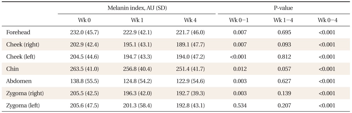
MI at the 1-week follow-up visit was significantly decreased compared to baseline on all sites, except the Lt. zygoma. MI at the 1-week follow-up visit on the abdomen, measured to minimize the effect of sunlight, was also decreased significantly compared to baseline. MI at the 4-week follow-up visit was not statistically significant when compared with MI at the 1-week follow-up visit. MI at the 4-week follow-up visit was significantly decreased compared to baseline on all 7 sites measured (Table 1).
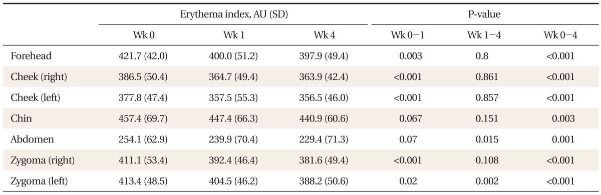
EI at the 1-week follow-up visit was significantly decreased compared to baseline on 5 sites, excluding the centre of the chin and the abdomen. EI at the 4-week follow-up visit was significantly decreased compared with the 1-week follow-up visit on the abdomen and Lt. zygoma. EI at the 4-week follow-up visit was significantly reduced compared to baseline on all 7 sites measured (Table 2).
DISCUSSION
The color of human skin is determined by many factors, including the quantity and chemical structure of its melanin pigments, non-melanin pigments such as Hb, and chemicals like lysogenic acid or liquorice.4) Our study showed that skin color changes occurred within 1 month after smoking cessation. MI reflected the melanin content of the skin and EI was related to the Hb content; both were significantly decreased at all sites measured within 1 month of stopping tobacco use.
Smoking induces oxidative stress, which has immunomodulatory effects by changing inflammatory cell function and releasing proteolytic enzymes.5) Accordingly, smoking tobacco is closely associated with numerous dermatologic conditions, including psoriasis, systemic lupus erythematosus, hair loss, hidradenitis suppurativa, genital warts, poor wound healing, wrinkling, and premature skin aging.6,7) Previous studies have reported that the frequency of oral mucosa pigmentation increases with the presence of smoking.8) In addition, a dose-response relationship was detected9) and disappearance of gingival pigmentation was observed following reduction in smoking.10)
Smoker's melanosis is hypothesized to occur as a result of the ability of nicotine to stimulate melanocyte activity and melanin production.11) The results of the current study, where MI decreased after smoking abstinence, are in line with previous observations.
Cigarette smoking seems to cause a generalized upward shift of the Hb distribution curve, resulting in a reduction in the ability of Hb to deliver oxygen to tissues. Nordenberg et al.12) confirmed that Hb levels are significantly higher in smokers. EI indicates Hb content that is closely related to skin blood flow,13) and EI was reduced in the current study; this suggests that there are changes of Hb and skin blood flow after smoking cessation. More research is needed to understand the underlying pathophysiologic mechanisms.
The most important strength of this study is that no other studies have reported skin color changes within 1 month of smoking abstinence. The main limitations of the present study are that all the subjects were male and the sample size was small. Furthermore, there is no comparison study.
Nevertheless, our study seemed to be meaningful because the 3 persons who constantly smoked the same amount during this study did not show any changes in MI and EI for a month, even if the sample size was small (3 persons were not included in 34 subjects). We did not adjust for any potential modifying effects of sunlight on skin color. However, our study was conducted from March to July, the spring to early summer in Korea, which is a period of very heavy sunlight. Generally sunlight is the one of factors which increases MI and EI. Nevertheless, our study clearly showed MI and EI decreases after smoking cessation.
The present study is the first to demonstrate changes in skin color within 1 month of smoking cessation. Additional investigations involving a pathological approach, both female and male subjects, and a larger sample size are now required. Visual impressions created by skin color, especially facial skin color, are important in interactions within and between human communities. We observed skin color changes after smoking cessation within a short time period. Skin color was measured through simple instrumental evaluation. Therefore, we anticipate that non-invasive instrumental evaluation of desirable effects on skin color after smoking cessation will play a positive role in maintaining smoking abstinence in routine clinical practice.